
ANGIOGRAPHYANDINTERVENTION.COM
Central Venous Access Devices


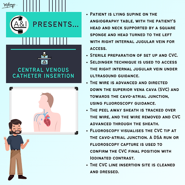
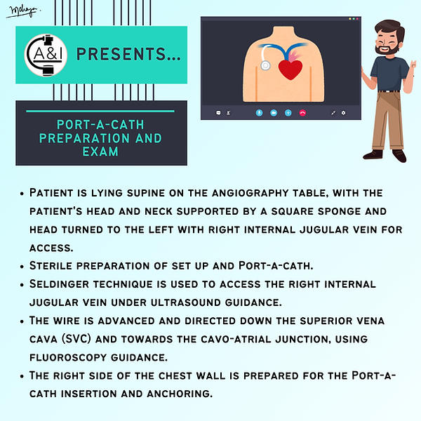
A&I Protocol


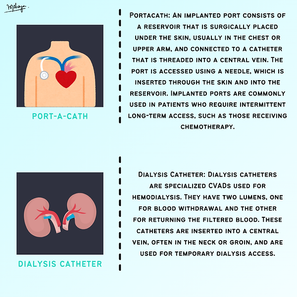
Common central venous access devices including PICC lines, Portacath and CVC.

DSA or Portacathogram showing the flow of contrast through the Portacath and into the right atrium of the heart.

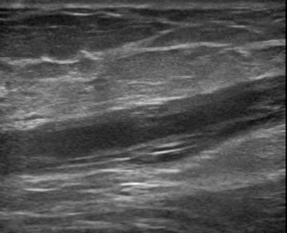
(Left) Transverse ultrasound showing the triad for PICC line access: brachial nerve bundle, brachial artery (pulsatile), and brachial vein underneath (compressible using the probe).
(Right) Longitudinal ultrasound of a basilic vein for PICC line puncture and access.
References
-
Lockwood J, Desai N. Central venous access. Br J Hosp Med (Lond). 2019 Aug 2;80(8):C114–9.
-
Saugel B, Scheeren TWL, Teboul JL. Ultrasound-guided central venous catheter placement: a structured review and recommendations for clinical practice. Crit Care. 2017 Aug 28;21(1):225.
-
Wadełek J. Haemodialysis catheters. Anestezjol Intens Ter. 2010;42(4):213–7.
-
Sansivero GE. Features and selection of vascular access devices. Semin Oncol Nurs. 2010 May;26(2):88–101.
-
Moureau N, Chopra V. Indications for peripheral, midline and central catheters: summary of the MAGIC recommendations. Br J Nurs. 2016 May 28;25(8):S15-24.